


Fungal infections of nails are a serious medical and social problem.
The pathogen is stable in the external environment and is quite easily transmitted from person to person.
If you detect the disease in time, you can protect yourself, your family members and others from infection.
And to understand when consultation with a qualified physician is necessary, it doesn’t hurt to know what nail fungus looks like.
There are many varieties of fungi.

And many of them can cause disease on the nails.
The general name of this unpleasant sore is onychomycosis.
Depending on which particular fungus led to the development of onychomycosis, the disease is called.
What does nail fungus look like: types of disease
The most common are:
- Athlete's foot.
- Rubrophytia.
- Trichophytosis.
- Favus.
- Candidiasis.
The causative agents of each of these diseases affect not only nails, but also skin and hair.
Therefore, if any changes appear on the body, even minor ones at first glance, it is better to see a doctor.
Perhaps this will help identify the onset of the disease and stop the spread of infection.
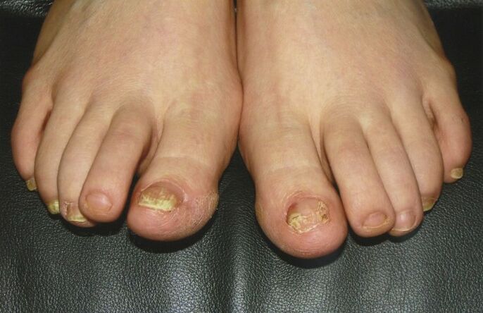
Athlete's foot
Interestingly, when the disease occurs on the nails, the first and fifth toes are most often affected.
Why the fungus chooses them is unknown.
First of all, the appearance of the nail changes:
- The pink color gives way to yellowish.
- The surface becomes dull and loses its healthy shine.
- Thickenings and tubercles appear on the plate.
- Dense growths (hyperkeratosis) develop under it.
- Canary-colored spots or stripes appear in the thickness of the nail.
The shape of the nail itself lasts quite a long time - several weeks or even months.
The free edge is gradually destroyed.
It becomes as if corroded, uneven.
Another feature of athlete's foot is that the disease develops only on the feet.
The same picture as toenail fungus looks like on the hands will not be the same.
If so, it is not athlete's foot.
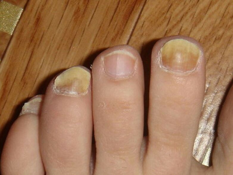
Rubrophytia
In this case, onychomycosis is caused by a fungus called trichophyton red.
There are three forms of the disease:
- Normotrophic form.
- Hypertrophic.
- Atrophic variety.
In the normotrophic form of onychomycosis, the nail plate does not collapse for a long time.
White or yellow stripes (leukonychia) appear in its thickness.
At first they are separated from each other, but gradually merge into a single spot.
In the typical course of the disease, the border at the base of the nail remains unchanged.
Hypertrophic nail rubrophytosis proceeds differently.
The record first becomes dull and loses its shine.
It thickens due to growths (hyperkeratosis) underneath.
The nail takes on a beak-like shape and crumbles easily.
And this applies not only to the free edge.
The long course of the disease gives the nails a resemblance to bird claws - onychogryphosis.
The atrophic form is also special.
The nail becomes dull and becomes dirty gray in color.
In a fairly short time, the nail plate becomes thinner and destroyed.
Along the edges, at the nail fold, the nail tissue remains, but it may also disappear.
Trichophytosis
This fungus causes disease of the entire surface of the skin.
Onychomycosis develops only in half of the patients, and the nails of the hands are affected.
What the initial stage of nail fungus looks like with trichophytosis does not allow making an accurate diagnosis, since there are similarities with other mycoses.
The surface of the nail plate becomes dull and the color becomes gray.
Over time, the nail develops fragility and crumbles.
In some cases, it may even peel off from its bed.
The process is lengthy and can take several years.
Favus
A synonym for this pathology is scab.
Children rarely get sick.
The disease has a long, chronic course.
The causative agents are several fungi from the genus Trichophyton.
First, due to subungual keratosis, the plate thickens and moves away from the nail bed.
At the same time, it begins to crumble.
In its thickness one can observe a single yellow spot - the scutula.
Gradually it acquires a dirty color.
Death of the nail occurs after several months from the onset of the disease.
The process is usually accompanied by other manifestations of the hair and skin.
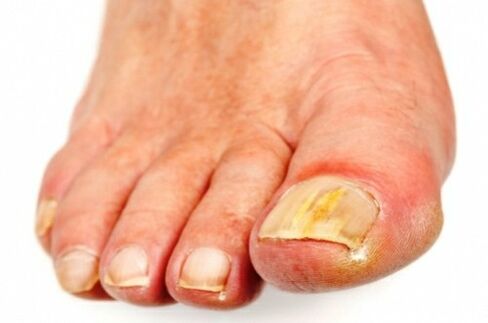
Candidiasis
Yeast fungi, the causative agent of this infection, normally live on the human body and on mucous membranes.
Activation of the infection leads to the appearance of systemic lesions, which can also spread to the nails.
The reasons for this may be
- I. Uncontrolled use of antibiotics.
- II.Immunodeficiency states.
- III. Taking hormonal medications.
- IV. Treatment with cytostatics.
- V. Hypovitaminosis.
The hands and feet are affected with equal frequency.
Visually, what toenail fungus looks like in the initial stage with a yeast infection is difficult to confuse with other diseases.
The nail plate acquires a brown color and becomes bumpy due to stripes and depressions.
Whitish spots appear on its surface and in its thickness.
They are loose and can be easily removed from the nail (if they are located superficially).
The nail itself delaminates, peeling away from the nail bed.
Along its edges, in the cuticle area, cheesy layers also appear.
It becomes red and inflamed.
The interdigital spaces are also affected.